

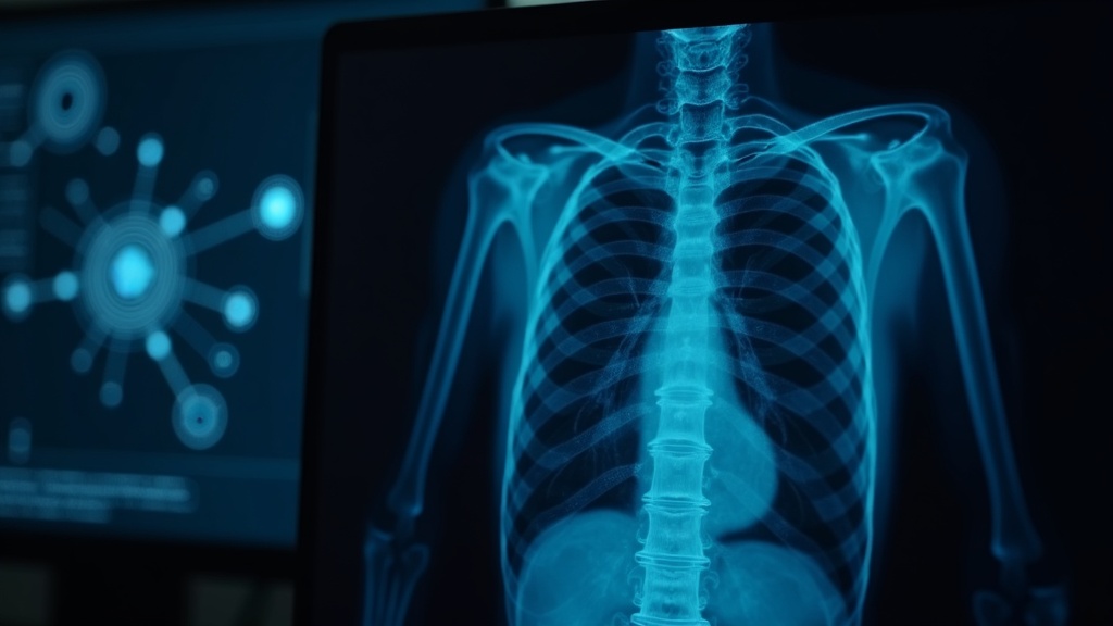
X-ray imaging has played a pretty big role in healthcare for decades, letting doctors spot broken bones, lung infections, and lots of other problems. Recently, artificial intelligence (AI) has been making waves in how these images are analyzed and interpreted. AI helps speed up diagnosis, give a boost to accuracy, and even catch subtle changes that human eyes might miss. In this article, I’ll walk you through how AI applications are changing the world of X-ray image processing, with practical tips and insights if you’re curious about how this technology works in the real world.
Why AI in X-Ray Image Processing Matters
Medical X-rays have always been important, but reading them accurately isn’t always easy. Even skilled radiologists can get tired, and subtle patterns might get missed, especially during busy shifts. With the rise of machine learning (ML) and deep learning, AI systems are now able to look at thousands of X-ray images and spot clues that would take humans much longer to process. That’s great for early detection and also helps reduce diagnostic errors.
The use of AI in radiology is growing quickly. According to market research from MarketsandMarkets, the AI-based medical imaging market is expected to reach over $2 billion in the next few years. Big hospitals are already investing in these tools, and smaller clinics are starting to follow suit as the software becomes more accessible. It’s clear that AI is set to play a key role in shaping the next stage of routine medical imaging.
Common AI Techniques Used for X-Ray Image Analysis
AI systems used in X-ray imaging rely mostly on machine learning techniques. Here’s a quick rundown of some terms and how they work in practice:
- Deep Learning: These models, especially convolutional neural networks (CNNs), can analyze pixellevel details in X-rays. CNNs have become the goto for detecting conditions like pneumonia, lung nodules, fractures, and more.
- Segmentation Algorithms: AI can automatically outline or highlight regions of interest in an X-ray, like a suspicious mass or a fractured bone. This helps radiologists zoom in on the most relevant areas.
- Image Enhancement: AI can improve image quality by reducing noise, clarifying boundaries, and highlighting abnormalities. This makes it easier to interpret challenging scans.
- Classification Tools: Some AI apps sort X-rays into categories (like “normal” or “abnormal”) before a human even looks at them, helping to speed up the triage process and focus attention where it matters most.
Along with these core techniques, some AI models use natural language processing to organize and extract findings from radiology reports, creating a loop between text and image interpretation. This kind of data blending is opening up new doors for pattern recognition and workflow automation in busy radiology departments.
Getting Started With AI-Boosted X-Ray Workflows
A lot of clinics and radiology departments are exploring AI applications for X-ray analysis. Making this transition smooth can be easier with a bit of planning. Here are some steps to help you get started:
- Choose The Right AI Tools: Start with user-friendly software that works with your existing imaging equipment. Some tools are cloud-based and require no extra hardware, while others connect directly into PACS (picture archiving and communication systems).
- Train Your Staff: AI works best when radiologists and technicians understand how it fits into their workflow. Spend some time on hands-on training, so staff feel confident using the new system.
- Start With Pilot Projects: Rather than going all in right away, try a pilot phase focused on a specific condition or set of exams. This approach makes it easier to measure improvements and spot any bumps early.
- Monitor Performance: Keep track of accuracy, turnaround time, and how doctors and patients are responding. Feedback helps you fine-tune your system and decide what to roll out next.
- Integrate With Care Pathways: The biggest benefit comes when AI findings are connected directly to clinical decisions. Set up clear processes for how and when these insights are used in patient care.

Things to Know Before Implementing AI in X-Ray Imaging
Using AI for X-ray image processing has its own challenges. Here are some points worth paying attention to, and tips for making the most of these tools:
- Data Privacy: Medical images contain sensitive information. Make sure any AI vendor you work with follows privacy standards like HIPAA or GDPR, and ask how your data will be protected.
- Data Quality: AI models work best with clear, welllabeled images. Make sure your imaging processes are consistent and that any data you feed into an algorithm is as clean as possible.
- Bias and Accuracy: AI learns from past data. If your historical data had biases (like fewer images from certain patient groups), the model might pick those up. It’s smart to check regularly for any fairness issues.
- Human Oversight: AI is a support tool and not a replacement for experienced radiologists. Always get a human to review the system’s suggestions and make final decisions, especially for complicated cases.
- Regulatory Compliance: In the U.S., the FDA reviews and clears many AI-powered medical tools. Only use software that’s been approved or cleared in your region.
Data Privacy
Patient privacy is a big deal. Be sure your AI platform offers features like data encryption and anonymization. Many vendors also offer onpremises deployments so that data never has to leave your facility. It’s also wise to check in with IT about hosting arrangements when choosing any cloud solution.
Data Quality
Low-quality or mislabeled X-rays can really throw off AI predictions. Some clinics set up regular “data hygiene” checks to make sure images are clear, consistent, and labeled accurately before they’re added to any training set. Creating a brief checklist for quality before adding new images to your system can help avoid mistakes down the road.
Bias and Accuracy
Diverse and representative data helps prevent algorithm bias. Some hospitals team up with universities or use open datasets to widen the range of cases the model sees during training. Periodic audits for model performance can reveal if specific patient groups are being underserved.
Human Oversight
Even when AI is right 99% of the time, that remaining 1% can be important. Training radiologists to review AI-generated results helps catch unusual situations and reinforces trust in the system. Some departments regularly schedule review meetings to double down on staff training and encourage a culture of shared learning.
Regulatory Compliance
Check for FDA clearance or your country’s regulatory body approval before adopting any clinical AI. The latest approved devices are listed on the FDA’s database. Local regulations might vary, so always ask your vendor for documentation to prove compliance with your medical environment.
Rolling out AI responsibly makes a big difference in building confidence and getting the most out of your investment. Take your time during the implementation phase, and don’t hesitate to reach out to vendors and other healthcare facilities that have already made the move for real-world advice.
Advanced Tips and Trends in AI-Powered X-Ray Analysis
If you’re ready to try out more advanced applications, these tips might come in handy:
Keep Training the AI Model: The more data you feed into the system (with the right checks, of course), the better it can spot new problems or rare conditions. Collecting fresh data regularly is key to keeping the tool up to date. Listen to staff feedback as well—sometimes new use cases come from daily routines.
Integrate Multiple Modalities: Some AI platforms combine X-ray data with CT, MRI, or basic patient info, which can help doctors make more tailored decisions. When images and medical histories are put together, doctors can get a fuller picture of what’s happening with the patient.
Use AI for Workflow Automation: AI can sort, flag, and even prioritize studies based on urgency. This is especially helpful in busy emergency departments where getting results faster can change patient outcomes. Over time, AI might even be able to suggest follow-up tests or provide risk scores to support clinical pathways.
Tap Into Telemedicine: Clinics working with remote radiologists can speed up second-opinion reviews by using AI to auto-highlight suspicious images before they reach a human expert. This not only adds to efficiency but also helps support facilities. It’s a solid way for small clinics to scale up expertise without hiring extra staff.
Keeping up with the newest trends and periodically updating your AI tools is an easy way to keep getting value from your investment. Staying up to date can unlock better workflows and better results for your patients—so don’t be afraid to experiment and keep learning.

Real-World Impact of AI in X-Ray Processing
AI-powered X-ray analysis isn’t just some futuristic idea. Hospitals and clinics around the world are already seeing the benefits. For example, at Mount Sinai Hospital in New York, AI systems help flag cases of potential pneumonia and alert doctors quickly so those patients get attention sooner (source).
Community hospitals have managed to improve their patient flow by using AI triage systems, cutting wait times, and reducing unnecessary followup scans. In some busy healthcare systems, AI has helped lower error rates by acting as a second set of eyes, which boosts confidence for doctors and patients alike.
- Lung Disease Detection: AI is often put to work to spot early signs of tuberculosis, pneumonia, or lung nodules in chest X-rays.
- Bone Fracture Identification: Algorithms can flag possible fractures or misalignments, sometimes catching injuries that could go unnoticed on a rushed shift.
- Cancer Screening: Automated tools can scan for tumors or areas of abnormal tissue in mammography or chest images, supporting early intervention and timely treatment.
Not only does this technology step up the speed of diagnosis, but it also adds a safety net by reducing the odds of missing critical findings. As more clinics share their results, it’s likely we’ll keep seeing growth in AI’s role, and more examples will pop up from all over the healthcare map.
Frequently Asked Questions
Here are some questions I get from other healthcare pros and anyone wondering about AI in X-ray imaging:
Question: Does AI replace radiologists?
Answer: No, AI is used mostly as an assist tool. It can help scan large numbers of images quickly and flag up problem areas, but final interpretation is still up to a human radiologist.
Question: How accurate are AI systems with X-rays?
Answer: In studies, some AI models match or even beat human radiologists at specific tasks (like lung nodule detection), but results can vary based on data quality and the problem type. Human review is always recommended to double-check the system’s findings.
Question: Can small hospitals use AI for X-ray analysis?
Answer: Yes, plenty of cloudbased platforms offer payperuse models that are affordable for even small clinics. Some systems can work directly with existing digital X-ray equipment and don’t require massive investments.
Final Thoughts
AI in X-ray image processing is giving healthcare teams a fresh set of tools for faster, more reliable diagnosis. By blending smart algorithms with human expertise, clinics can spot diseases earlier, improve patient care, and reduce burnout for radiologists. The tech is growing fast, so staying up to date and asking good questions when picking a platform will keep you at the leading edge. If you’re thinking about checking out AI for X-ray workflow, now is a great time to dig in and see what’s out there.